Encephalitis > Volume 1(4); 2021 > Article
Yoon, Kim, Ahn, and Chu: Progressive multifocal leukoencephalopathy successfully treated with mefloquine and literature review

Abstract

Progressive multifocal leukoencephalopathy (PML) is an opportunistic infection due to reactivation of John Cunningham virus (JCV). The diagnosis depends on evidence from clinical, imaging, and virologic studies. When the cerebrospinal fluid shows a negative polymerase chain reaction result, brain biopsy is required to confirm the diagnosis. PML has no standard treatment except for immune reconstitution. The anti-JCV effect of mefloquine, however, is supported by some studies, and if brain biopsy is difficult, a mefloquine trial can be considered. We describe a case of possible PML successfully treated with mefloquine.

Introduction

Progressive multifocal leukoencephalopathy (PML) is a rare fatal infection caused by John Cunningham virus (JCV) that usually occurs in immunocompromised patients. Since any area of the brain can be involved, PML shows diverse clinical features. The diagnostic criteria of PML are comprised of evidence from clinical, imaging, and virologic studies [1].
PML has no approved treatment except for immune reconstitution. Although in vitro studies and some case reports suggest an anti-JCV property of mefloquine [2-22], the efficacy of mefloquine for JCV is controversial [23,24]. We describe a possible PML case with negative cerebrospinal fluid (CSF) polymerase chain reaction (PCR) for JCV that was successfully treated with mefloquine.

Case Report

A 52-year-old male patient visited the emergency department with a 2-week history of positional vertigo, a 3-day history of tinnitus, ear pressure, and hearing loss in the left ear. He denied other neurologic symptoms. He had a history of coronary stent insertion for myocardial infarction and living-donor kidney transplantation for end-stage renal disease of unknown etiology. He was taking prednisolone 5 mg daily with mycophenolate mofetil 360 mg twice a day.
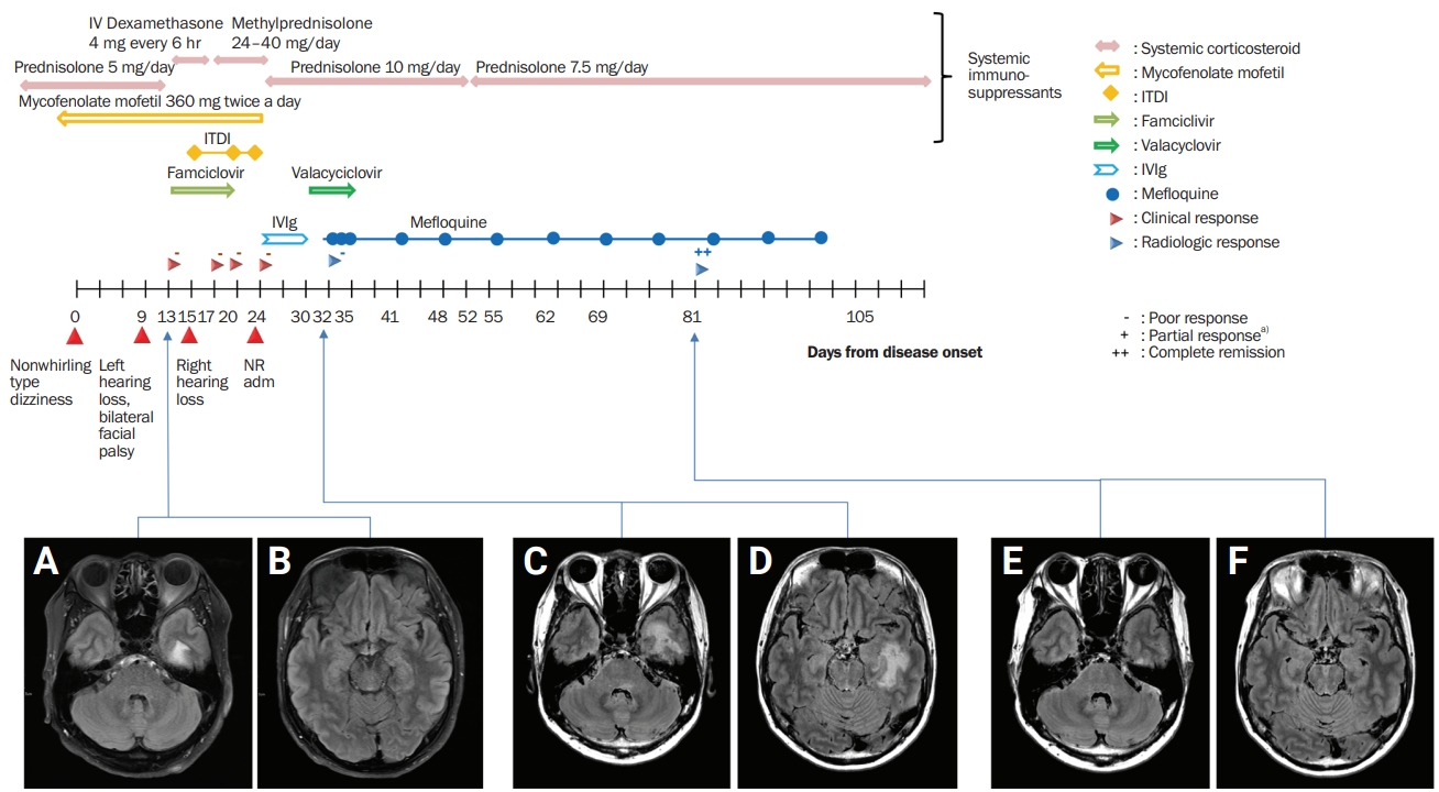
Neurologic examination revealed Weber lateralization to the right side, bilateral nasolabial fold blunting, and truncal ataxia. However, other cranial nerves, motor, sensory, and cerebellar function tests, and the otoscope examination were unremarkable. The initial impression was multiple cranial nerve palsy. Brain magnetic resonance imaging (MRI) revealed bilateral facial nerve enhancement (left > right) and subtle fluid-attenuated inversion recovery (FLAIR) hyperintensity and T1 hypointensity in the left temporal white matter (Figure 1A and B).
He was admitted to the otorhinolaryngology department with the initial impression of Ramsay-Hunt syndrome. The patient received intravenous dexamethasone 4 mg every 6 hours for 4 days, oral methylprednisolone 40 mg for 10 days, and famciclovir 500 mg for 7 days. Intratympanic dexamethasone was administered three times in the left ear and twice in the right ear, but the administration aggravated the patient’s bilateral sensorineural hearing loss.
He was referred to the neurology department and was admitted with the impression of encephalitis of infectious or autoimmune etiology. CSF examination showed marginal pleocytosis (leukocytes, 8 cells/µL; lymphocytes, 75%; others, 25%) with a normal protein level (39 mg/dL) and negative results for the Gram stain, bacterial culture, and cryptococcal antigen (Table 1). Valacyclovir 1,000 mg twice a day for 1 week and intravenous immunoglobulin G (IgG) 2 g/kg over 5 days were prescribed. The corticosteroid dose was tapered to oral prednisolone 10 mg daily. CSF PCR revealed a positive result for Epstein-Barr virus (EBV) and negative results for mycobacteria, cytomegalovirus, herpes simplex virus, varicella zoster virus (VZV), and JCV. The result of serum JCV PCR was positive. EBV viral capsid antigen (VCA) IgG was positive, but EBV VCA IgM was negative. Early antigen and EBV nuclear antigen tests were not conducted (Table 1).
After 2 weeks of antiviral treatment, follow-up brain MRI showed FLAIR hyperintensity and T1 hypointensity in the subcortical white matter of the left temporal lobe and dorsal pons (Figure 1C and D). The imaging findings suggested lymphoproliferative disease or PML. Whole-body positron emission tomography was performed to rule out the possibility of lymphoproliferative disease and revealed no abnormal hypermetabolism, which would suggest malignancy. In the meantime, an initial workup that included ganglioside antibodies, antinuclear antibody, antineutrophil cytoplasmic antibody, angiotensin-converting enzyme level, aquaporin-4 IgG, myelin oligodendrocyte glycoprotein IgG, vitamin B12, and folate level showed that these levels were normal. He was diagnosed with possible PML. Because of the patient’s poor response to the antiviral agent and the results of his extensive diagnostic workup, we excluded other differential diagnoses, including VZV leukoencephalopathy, central nervous system (CNS) vasculitis, and lymphoproliferative diseases. Nevertheless, the corticosteroid was tapered to prednisolone 10 mg equivalent, brain MRI showed increased extent of the FLAIR hyperintensity lesion, and pure-tone audiometry (PTA) showed progressive bilateral sensorineural hearing loss. Since steroid tapering was not sufficient to halt disease progression, mefloquine 250 mg/day for 3 days was introduced and maintained at 250 mg every week. After mefloquine treatment, his neurologic deterioration stopped, and no other focal neurologic deficit, other than the presenting sensorineural hearing loss, appeared. In the 6-week follow-up MRI, previous white matter lesions were markedly decreased, and serum JCV PCR was negatively reversed (Figure 1E and F). Initially, we planned maintenance treatment with mefloquine to continue until radiologic remission or confirmed recovery of hearing. However, the maintenance therapy of mefloquine 250 mg/week was administered for only 8 weeks due to the patient’s refusal. At his nadir, he was only able to follow simple commands, but after treatment, his sensory aphasia improved to almost normal and his cognitive function improved to independent activities of daily living. A change in his hearing deficit, however, was not assessed due to the patient’s refusal for PTA follow-up.

Discussion

PML is prevalent in immunocompromised patients. Although JCV infects more than 50% of the adult population, its replication is suppressed by antigen-specific T cells in immune-competent individuals [1]. In an immune-compromised subject, however, JCV can replicate in oligodendrocytes and astrocytes causing lytic necrosis, which is a key factor in the pathophysiology of PML [25].
No single criterion has been established for the diagnosis of PML. The American Academy of Neurology suggests that clinicians diagnose PML based on evidence from clinical, neuroimaging, and virologic studies [1]. Given these criteria, the definite or probable PML case requires positive CSF JCV PCR or histopathologic evidence in brain biopsy. However, several reports describe negative CSF JCV PCR cases that were finally diagnosed as PML [3,22,26-34]. In a search on PubMed, at least 11 case reports published in English were available, and each case was biopsy-proven [3,22,26-34] (Table 2). Some new diagnostic criteria of PML have been proposed to overcome this limitation [35]. Our case had clinical and imaging features supporting PML and was categorized as possible PML. Other possibilities, like CNS vasculitis and VZV leukoencephalopathy, were excluded through an extensive diagnostic workup. A brain biopsy was required to confirm the diagnosis of PML; nonetheless, brain biopsy was spared in this patient due to its invasiveness and empirically treated as PML.
The only approved treatment of PML is immune reconstruction. This approach is based on the fact that PML is one of opportunity infections. The removal of immunosuppressants in treatment-related PML and antiretroviral therapy in human immunodeficiency virus-associated PML are good examples. Treating PML with immune checkpoint inhibitor is also a similar strategy [36].
Other strategies are based upon in vitro studies. Mirtazapine or atypical antipsychotics were expected to inhibit viral entry into cells blocking 5HT2A receptors, which is a cellular receptor for JCV. For these medications to be validated as treatment options for PML, their toxicity should be tolerable in the therapeutic range, and the drugs should be delivered to the CNS. In vitro studies suggest that mefloquine not only has an anti-JCV property by inhibiting viral DNA replication but also sufficiently penetrates the blood brain barrier [2]. Moreover, these in vitro studies are supported by several case reports of PML successfully treated with mefloquine [3-22,37]. In PubMed, at least 21 case reports published in English were available, and these cases even included treatment without additional immune reconstitution therapy [3-22,37] (Table 3). Some clinical studies failed to show the clinical efficacy of mefloquine [23], but some points must be considered. A large clinical trial of PML is difficult due to its rarity. In addition, it seems that ABCB1/MDR1 gene polymorphism has an important role in pharmacokinetics and efficacy [24], contributing to the negative results of the trial [23].
In the present case, although the virologic evidence was not fulfilled, clinical and imaging findings led to the impression of PML. Immunosuppressants were tapered but failed to halt the disease progression, and mefloquine treatment was administered.
According to pharmacokinetic studies, the biologic half-life of mycophenolate, prednisolone/methylprednisolone, and intravenous Ig were reported as 9 to 17 hours, 12 to 36 hours, and 14 to 35 days, respectively [38-40]. Since 4 half-lives is usually considered sufficient time to reach the steady state, it seems that the half-lives of the corticosteroid and mycophenolate mofetil are too short and the half-life of intravenous Ig is too long to explain our patient’s delayed and prolonged treatment response 1 month after onset. Therefore, with all these confounding factors including other immune-related medication changes, it is reasonable to conclude that mefloquine led to improvement of the PML.
Therefore, physicians can learn two points from this case. A negative CSF JCV PCR does not always rule out PML. Moreover, when PML is clinically highly suspicious and brain biopsy is difficult, a mefloquine trial can be considered as an option.

Notes

Conflicts of Interest

Kon Chu has been on the editorial board of encephalitis since October 2020. He was not involved in the review process of this case report. No other potential conflict of interest relevant to this article was reported.

Author Contributions

Conceptualization: Chu K; Data curation: Yoon S, Chu K; Formal analysis: Yoon S, Chu K; Investigation: Ahn SJ, Kim Y; Writing–original draft: Yoon S; Writing–review and editing: all authors.

Figure 1.

Hyperintensity in the subcortical white matter was seen in the fluid-attenuated inversion recovery sequence of the brain magnetic resonance imaging.

Left inferior temporal gyrus (A) and left superior temporal gyrus (B) on the initial image; and left inferior (C) and superior temporal gyrus and left dorsal pons (D) before mefloquine treatment. Decreased hyperintensity in the left temporal areas (E, inferior; F, superior) after mefloquine treatment.
IV, intravenous; ITDI, intratympanic dexamethasone injection; IVIg, IV immunoglobulin G.
a)Clinical improvement with remnant neurologic deficit.
encephalitis-2021-00094f1.jpg
Table 1
Laboratory results of blood and cerebrospinal fluid of the patient
Lab finding Initial visit Second admission 6 wk after mefloquine Normal range
Blood
 White blood cell (/μL) 4.78 8.51 7.98 4–10
 Hemoglobin (mg/dL) 9 8.3 13.8 12–16
 Platelets (×103/μL) 214 196 365 130–400
 Absolute neutrophil count (/μL) 3824 7829 1.3 1–5
 C-reactive protein (mg/L) 0.06 0.01 0.02 0–0.5
 Sodium (mEq/L) 138 139 133 135–145
 Potassium (mEq/L) 4.2 4.5 3.8 3.5–5.5
 Albumin (g/dL) 3.6 3.0 4.4 3.3–5.2
 Glucose (mg/dL) 216 70–110
 EBV PCR Positive NA
 JCV PCR Positive Negative
 VZV PCR Negative NA
Cerebrospinal fluid
 Cell count (/μL) NA 8 NA 0–5
 Polymorphonuclear cell (%) 0
 Lymphocytes (%) 75
 Other cells (%) 25
 Protein (mg/dL) 39 15–45
 Glucose (mg/dL) 69 40–70
 FTA-ABS Nonreactive Nonreactive
 JCV PCR Negative Negative
 EBV PCR Positive Negative

EBV, Ebstein-Barr virus; PCR, polymerase chain reaction; NA, not available; JCV, John Cunningham virus; VZV, varicella zoster virus; FTA-ABS, fluorescent treponemal antibody absorption.

Table 2
Summary of case report of progressive multifocal leukoencephalopathy with false-negative PCR in CSF
Study Age (yr)/sex Presenting symptom Comorbidity Treatment MRI finding CSF study Brain biopsy
Kuhle et al., 2011 [26] 48/F L side hypesthesia and dysesthesia RRMS Prednisolone Compatible with MS Negative PCR for JCV Polyomavirus particles on EM
Nonenhancing and faintly enhancing ribbon-like lesion
Silverio et al., 2015 [22] 69/M Progressive dysarthria and R hemiparesis Follicular lymphoma Chemotherapy Multiple confluent foci of FLAIR hyperintensity involving the inferior R and L frontal lobes, as well as periventricular regions Negative PCR for JCV Chromatin margination and viropathic change within oligodendrocytes
Babi et al., 2015 [27] 75/F Progressive L hemiplegia and global decline Rheumatoid arthritis Methotrexate, adalimumab Asymmetric subcortical FLAIR HIS involving R frontoparietal subcortical WM Negative PCR for JCV Viral inclusion in enlarged oligodendroglial nucleus
Lee et al., 2019 [28] 44/M Dysphagia, memory disturbance, Seizure AIDS HAART Multifocal patchy lesions involving subcortical region of both frontal, R temporoparietal, L thalamus, striatocapsular regions Negative PCR for JCV Large infected oligodendrocytes with inclusion-bearing dark nuclei
High JCV DNA titer of brain biopsy specimen
van der Kolk et al., 2016 [29] 49/M Aphasia, dyscalculia, hyperesthesia of the R arm, and headache NA NA Large confluating asymmetric white matter hyperintensities lesions in the frontal and parietal lobes Negative PCR for JCV Positive PCR for JCV on the biopsy material
Kharfan-Dabaja et al., 2007 [30] 51/M Confusion and disorientation, dysnomia and progressive R upper extremity weakness → seizure → receptive aphasia, R hemiparesis, and cortical blindness Follicular NHL and secondary myelodysplasia GVHD prophylaxis with methotrexate , tacrolimus, alloHCT T2 hyperintensity in periventricular white matter clustered within the L centrum semiovale Negative PCR for JCV Extensive demyelination, presence of naked axons, reactive gliosis, and lipid-laden macrophages
Occasional nuclei with a basophilic ground glass appearance, suggestive of inclusions
The presence of viral particles typical of the papovavirus group in multiple cells in EM
Chowdhary and Chamberlain 2008 [31] 51/M Progressive confusion, dysarthria, and visual disturbance Myelodysplasia and NHL Allogenic bone marrow transplantation Multifocal T2HSI lesions including L frontal, parietal, and occipital lobes Negative PCR for JCV for twice Multiple enlarged, basophilic nuclei of infected oligodendrocytes intranuclear accumulation of spherical and filamentous viral particles typical of the papovavirus group
Vidarsson et al., 2002 [32] 63/M Progressive memory loss and R visual disturbances Follicular lymphoma Fludarabine, mitoxantrone, dexamethasone Multifocal T2HSI lesion in L parieto-occipital area Negative PCR for JCV Abnormal astrocytes with hyperchromaticnuclei, and oligodendrocytes with enlarged nuclei and “ground glass”' appearance
Landry et al. 2008  [33] 31/F L facial palsy and L sided weakness Job’s syndrome (HIES) IVIg Atypical T2HSI pattern Negative PCR for JCV Demyelination, myelin-debris-laden foamy macrophages, enlarged nuclei but no definitive intranuclear inclusions in oligodendroglial cells and no bizarre astrocytes
Multiple sclerosis treated with high-dose corticosteroid and plasma exchange Polyomavirus particles in EM finding
Sikkema et al., 2013 [34] 74/F Progressive symptoms of motor imbalance, fatigue, weight loss, and impaired cognitive function DLBCL RCHOP T2HSI lesions in L thalamus/mesencephalon, R subcortical frontal lobe Negative PCR for JCV Reactive gliosis and in the middle of a cell with a viral nuclear inclusion
Garrote et al., 2015 [3] 50/M Visual disturbance, diminished muscular strength in the R arm and vesicular-papular lesions in the L ophthalmic branch region of the V cranial nerve Chronic lymphocytic leukemia   Fludarabine, cyclophosphamide and rituximab T2 hyperintensity in bilateral parietal and occipital lobules including internal capsule Negative PCR for JCV Infiltration of the brain tissue by foamy macrophages and mature lymphocytes with perivascular clustering
Loss of myelin in immunohistochemistry
Reactive astrocytes with polymorphic nuclei and prominent nucleoli

PCR, polymerase chain reaction; CSF, cerebrospinal fluid; MRI, magnetic resonance imaging; F, female; M, male; R, right; L, left; RRMS, relapse and remitting multiple sclerosis; MS, multiple sclerosis; JCV, JCV, John Cunningham virus; EM, electron microscopy; FLAIR, fluid-attenuated inversion recovery; HIS, high signal intensity; AIDS, acquired immune deficiency syndrome; HAART, highly active antiretroviral therapy; NA; not available; NHL, non-Hodgkin lymphoma; GVHD, graft versus host disease; alloHCT, allogeneic hematopoietic cell transplantation; BMT, bone marrow transplantation; HIES, hyper immunoglobulin E (IgE) syndrome; IVIg, intravenous IgG; DLBCL, diffuse large B-cell lymphoma; RCHOP, rituximab, cyclophosphamide, hydroxydaunorubicin, oncovin, and prednisone.

Table 3
Summary of case report of PML successfully treated with mefloquine
Study Age (yr)/sex MRI lesion (T2/FLAIR hyperintensity) Lesion enhancement Comorbid diseases and treatment Treatment for PML Interval between the initial symptom onset and diagnosis (mo) Clinical outcome
Garrote et al., 2015 [3] 50/M Bilateral parietal and occipital lobules, and internal capsule - CLL treated with rituximab, fludarabine, cyclophosphamide Mefloquine and dexamethasone NA Marked improvement
Shin et al., 2014 [4] 67/M R Parietal lobe - IgAN on prednisone Mefloquine NA Marked improvement
Nishigori et al., 2019 [5] 68/M Bilateral MCPs, pons and cerebellum + RA treated with prednisolone, and methotrexate for 9 years Mefloquine and mirtazapine 6 Marked improvement
Hamaguchi et al., 2020 [6] 68/M R MCP and cerebellar hemisphere + RA, SLE on prednisolone, tacrolimus Mefloquine and mirtazapine 5 Marked improvement
Ishikawa et al., 2018 [7] 36/M Bilateral temporoparietal lobe - SLE, HLH on prednisolone, cyclosporin A, rituximab, cyclophosphamide, mycophenolate mofetil Mefloquine and mirtazapine 3 Marked improvement
Nambirajan et al., 2017 [8] 44/M Bilateral parieto-occipital subcortical and deep white matter - - Mefloquine and cotrimoxazole 1 Marked improvement
Gofton et al., 2011 [9] 54/F R cerebellum and brainstem - Sarcoidosis on steroid Mefloquine 6 Marked improvement
Hervás et al., 2015 [10] 51/M Bilateral MCPs and R frontal subcortical white matter + RRMS treated with natalizumab Intravenous methylprednisolone and mefloquine 1 Marked improvement
Young et al., 2012 [11] 57/M R BG, thalamus, R frontal WM + HIV on HAART Mefloquine 3 Marked improvement
39/M R frontoparietal subcortical white matter - HIV on HAART Mefloquine NA Marked improvement
Epperla et al., 2014 [12] 72/M L frontal lobe + CLL s/p splenectomy Mefloquine and mirtazapine 1 Marked improvement
Sanjo et al., 2016 [13] 53/M L frontal, parietotemporal and R parietal lobes + Follicular lymphoma treated with CCRT Mefloquine, risperidone, and cytarabine 1.5 Marked improvement
Yoshida et al., 2014 [14] 40/F R occipital and L frontal lobes NA GVHD treated with calcineurin inhibitor and steroid Mefloquine and mirtazapine NA Marked improvement
Yoshida et al., 2015 [15] 66/M L frontal lobe - Chronic hepatitis C after liver transplantation, GVHD on tacrolimus and rapamycin Mefloquine NA Marked improvement
Shirai et al., 2014 [16] 51/M L MCP and cerebellar lesion - Chronic hepatitis B with hepatocellular carcinoma  Mefloquine and methylprednisolone 3 Some improvement
66/F R frontal lobe - SLE, DM, SSc Mefloquine and mirtazapine 2 Some improvement
Hirayama et al., 2011 [17] 60/M Bilateral frontoparietal lobe - Sarcoidosis Mefloquine 4 Marked improvement
McGuire et al., 2011 [18] 74/F R frontal lobe + Idiopathic isolated CD8+ T-lymphocytopenia Mefloquine and mirtazapine NA Some improvement
Ishii et al., 2018 [19] 37/F Bilateral cerebral peduncles, internal capsule, corpus callosum, and deep white matter of the L frontal lobe and bilateral periventricular area - SLE treated with prednisolone, mycophenolate mofetil Mefloquine 5 Some improvement
Ikeda et al., 2017 [20] 32/F L frontal lobe NA SLE treated with oral prednisolone, tacrolimus and cyclophosphamide pulse Mefloquine and mirtazapine 1 Marked improvement
Nakayama et al., 2020 [21] 73/F L MCP, cerebellar hemisphere, brainstem NA ET treated with ruxolitinib Mefloquine and mirtazapine 7 Some improvement
Silverio et al., 2015 [22] 69/M L inferior frontal lobe to corona radiata - Follicular lymphoma treated by rituximab, Pulmonary sarcoidosis Mefloquine and mirtazapine 2 Some improvement
Kurmann et al., 2015 [37] 56/M L medial thalamus, hypothalamus, mesencephalon, and tegmentum pontis - CVID on IVIg Mefloquine and mirtazapine 2 Marked improvement

PML, progressive multifocal leukoencephalopathy; MRI, magnetic resonance imaging; FLAIR, fluid-attenuated inversion recovery; M, male; F, female; CLL, chronic lymphocytic leukemia; NA, not available; R, right; L, left; IgAN, immunoglobulin A nephropathy; MCP, middle cerebellar peduncle; RA, rheumatoid arthritis; SLE, systemic lupus erythematosus; HLH, hemophagocytic lymphohistiocytosis; RRMS, relapse and remitting multiple sclerosis; BG, basal ganglia; WM, white matter; HIV, human immunodeficiency virus; HAART, highly active antiretroviral therapy; s/p, status post operation; CCRT, concurrent chemoradiation therapy; GVHD, graft versus host disease; DM, dermatomyositis; SSc, systemic sclerosis; ET, essential thrombocytopenia; CVID, common variable immune deficiency; IVIg, intravenous immunoglobulin.

References

1. Berger JR, Aksamit AJ, Clifford DB, et al. PML diagnostic criteria: consensus statement from the AAN Neuroinfectious Disease Section. Neurology 2013;80:1430–1438.
crossref pmid pmc
2. Brickelmaier M, Lugovskoy A, Kartikeyan R, et al. Identification and characterization of mefloquine efficacy against JC virus in vitro. Antimicrob Agents Chemother 2009;53:1840–1849.
crossref pmid pmc
3. Garrote H, de la Fuente A, Oña R, et al. Long-term survival in a patient with progressive multifocal leukoencephalopathy after therapy with rituximab, fludarabine and cyclophosphamide for chronic lymphocytic leukemia. Exp Hematol Oncol 2015;4:8.
crossref pmid pmc
4. Shin JW, Jung KH, Lee ST, et al. Mefloquine improved progressive multifocal leukoencephalopathy in a patient with immunoglobulin A nephropathy. J Clin Neurosci 2014;21:1661–1664.
crossref pmid
5. Nishigori R, Warabi Y, Shishido-Hara Y, et al. Inflammatory cerebellar PML with a CD4/CD8 ratio of 2.9 showed a favorable prognosis in a patient with rheumatoid arthritis. Intern Med 2019;58:3323–3329.
crossref pmid pmc
6. Hamaguchi M, Suzuki K, Fujita H, et al. Successful treatment of non-HIV progressive multifocal leukoencephalopathy: case report and literature review. J Neurol 2020;267:731–738.
crossref pmid
7. Ishikawa Y, Kasuya T, Ishikawa J, Fujiwara M, Kita Y. A case of developing progressive multifocal leukoencephalopathy while using rituximab and mycophenolate mofetil in refractory systemic lupus erythematosus. Ther Clin Risk Manag 2018;14:1149–1153.
crossref pmid pmc
8. Nambirajan A, Suri V, Kataria V, Sharma MC, Goyal V. Progressive multifocal leukoencephalopathy in a 44-year old male with idiopathic CD4+ T-lymphocytopenia treated with mirtazapine and mefloquine. Neurol India 2017;65:1061–1064.
crossref pmid
9. Gofton TE, Al-Khotani A, O’Farrell B, Ang LC, McLachlan RS. Mefloquine in the treatment of progressive multifocal leukoencephalopathy. J Neurol Neurosurg Psychiatry 2011;82:452–455.
crossref pmid
10. Hervás JV, Presas-Rodríguez S, Crespo-Cuevas AM, et al. Progressive multifocal leukoencephalopathy associated to natalizumab extended dosing regimen. Neurodegener Dis Manag 2015;5:399–402.
crossref pmid
11. Young BE, Yeo TR, Lim HT, et al. Progressive multifocal leukoencephalopathy with immune reconstitution inflammatory syndrome (PML-IRIS): two case reports of successful treatment with mefloquine and a review of the literature. Ann Acad Med Singap 2012;41:620–624.
crossref pmid
12. Epperla N, Medina-Flores R, Mazza JJ, Yale SH. Mirtazapine and mefloquine therapy for non-AIDS-related progressive multifocal leukoencephalopathy. WMJ 2014;113:242–245.
pmid
13. Sanjo N, Kina S, Shishido-Hara Y, et al. Progressive multifocal leukoencephalopathy with balanced CD4/CD8 T-cell infiltration and good response to mefloquine treatment. Intern Med 2016;55:1631–1635.
crossref pmid
14. Yoshida H, Ohshima K, Toda J, et al. Significant improvement following combination treatment with mefloquine and mirtazapine in a patient with progressive multifocal leukoencephalopathy after allogeneic peripheral blood stem cell transplantation. Int J Hematol 2014;99:95–99.
crossref pmid
15. Yoshida T, Kawamoto M, Togo M, et al. Progressive multifocal leukoencephalopathy developing after liver transplantation showing marked neurological symptom improvement and arrest of further deterioration of imaging findings: a case report. J Neurol Sci 2015;359:1–3.
crossref pmid
16. Shirai S, Yabe I, Kano T, et al. Usefulness of 11C-methionine-positron emission tomography for the diagnosis of progressive multifocal leukoencephalopathy. J Neurol 2014;261:2314–2318.
crossref pmid
17. Hirayama M, Nosaki Y, Matsui K, et al. Efficacy of mefloquine to progressive multifocal leukoencephalopathy initially presented with parkinsonism. Clin Neurol Neurosurg 2012;114:728–731.
crossref pmid
18. McGuire JL, Fridman V, Wüthrich C, Koralnik IJ, Jacobs D. Progressive multifocal leukoencephalopathy associated with isolated CD8+ T-lymphocyte deficiency mimicking tumefactive MS. J Neurovirol 2011;17:500–503.
crossref pmid pmc
19. Ishii J, Shishido-Hara Y, Kawamoto M, et al. A punctate magnetic resonance imaging pattern in a patient with systemic lupus erythematosus is an early sign of progressive multifocal leukoencephalopathy: a clinicopathological study. Intern Med 2018;57:2727–2734.
crossref pmid pmc
20. Ikeda J, Matsushima A, Ishii W, et al. Brain biopsy is more reliable than the DNA test for JC virus in cerebrospinal fluid for the diagnosis of progressive multifocal leukoencephalopathy. Intern Med 2017;56:1231–1234.
crossref pmid pmc
21. Nakayama K, Nakamura M, Konishi A, et al. JC virus granule cell neuronopathy associated with Ruxolitinib: a case report and review of the literature. eNeurologicalSci 2020;21:100269.
crossref pmid pmc
22. Silverio KA, Patel SA. Progressive multifocal leukoencephalopathy with negative JC virus PCR following treatment of follicular lymphoma: implications for biologics in the era of targeted cancer therapy. Case Rep Oncol Med 2015;2015:534529.
crossref pmid pmc
23. Clifford DB, Nath A, Cinque P, et al. A study of mefloquine treatment for progressive multifocal leukoencephalopathy: results and exploration of predictors of PML outcomes. J Neurovirol 2013;19:351–358.
crossref pmid pmc
24. Kerb R, Fux R, Mörike K, et al. Pharmacogenetics of antimalarial drugs: effect on metabolism and transport. Lancet Infect Dis 2009;9:760–774.
crossref pmid
25. Tan CS, Koralnik IJ. Progressive multifocal leukoencephalopathy and other disorders caused by JC virus: clinical features and pathogenesis. Lancet Neurol 2010;9:425–437.
crossref pmid pmc
26. Kuhle J, Gosert R, Bühler R, et al. Management and outcome of CSF-JC virus PCR-negative PML in a natalizumab-treated patient with MS. Neurology 2011;77:2010–2016.
crossref pmid pmc
27. Babi MA, Pendlebury W, Braff S, Waheed W. JC Virus PCR detection is not infallible: a fulminant case of progressive multifocal leukoencephalopathy with false-negative cerebrospinal fluid studies despite progressive clinical course and radiological findings. Case Rep Neurol Med 2015;2015:643216.
crossref pmid pmc
28. Lee SY, Ko HC, Kim SI, Lee YS, Son BC. Progressive multifocal leukoencephalopathy diagnosed by brain biopsy, not by the DNA test for JC virus. Asian J Neurosurg 2019;14:240–244.
crossref pmid pmc
29. van der Kolk NM, Arts P, van Uden IW, et al. Progressive multifocal leukoencephalopathy in an immunocompetent patient. Ann Clin Transl Neurol 2016;3:226–232.
crossref pmid pmc
30. Kharfan-Dabaja MA, Ayala E, Greene J, Rojiani A, Murtagh FR, Anasetti C. Two cases of progressive multifocal leukoencephalopathy after allogeneic hematopoietic cell transplantation and a review of the literature. Bone Marrow Transplant 2007;39:101–107.
crossref pmid
31. Chowdhary S, Chamberlain M. A progressive neurologic disorder with multiple CNS lesions: a neuroimaging clinicopathologic correlation: progressive multifocal leukoencephalopathy (PML). J Neuroimaging 2008;18:340–344.
crossref pmid
32. Vidarsson B, Mosher DF, Salamat MS, Isaksson HJ, Onundarson PT. Progressive multifocal leukoencephalopathy after fludarabine therapy for low-grade lymphoproliferative disease. Am J Hematol 2002;70:51–54.
crossref pmid
33. Landry ML, Eid T, Bannykh S, Major E. False negative PCR despite high levels of JC virus DNA in spinal fluid: implications for diagnostic testing. J Clin Virol 2008;43:247–249.
crossref pmid pmc
34. Sikkema T, Schuiling WJ, Hoogendoorn M. Progressive multifocal leukoencephalopathy during treatment with rituximab and CHOP chemotherapy in a patient with a diffuse large B-cell lymphoma. BMJ Case Rep 2013;2013:bcr2012008142.
crossref pmid pmc
35. Wijburg MT, Warnke C, Killestein J, Wattjes MP. Application of “Mentzer’s PML case definition” to natalizumab-treated patients in the setting of strict MRI-based pharmacovigilance. J Neurol 2020;267:2599–2602.
crossref pmid
36. Cortese I, Muranski P, Enose-Akahata Y, et al. Pembrolizumab treatment for progressive multifocal leukoencephalopathy. N Engl J Med 2019;380:1597–1605.
crossref pmid
37. Kurmann R, Weisstanner C, Kardas P, et al. Progressive multifocal leukoencephalopathy in common variable immunodeficiency: mitigated course under mirtazapine and mefloquine. J Neurovirol 2015;21:694–701.
crossref pmid
38. Wexler P, Anderson B, de Peyster A, et al. Encyclopedia of toxicology. 2nd ed. New York: Elsevier; 2005.
39. Staatz CE, Tett SE. Clinical pharmacokinetics and pharmacodynamics of mycophenolate in solid organ transplant recipients. Clin Pharmacokinet 2007;46:13–58.
crossref pmid
40. Koleba T, Ensom MH. Pharmacokinetics of intravenous immunoglobulin: a systematic review. Pharmacotherapy 2006;26:813–827.
crossref pmid


ABOUT
BROWSE ARTICLES
EDITORIAL POLICY
FOR CONTRIBUTORS
Editorial Office
101, Daehak-ro, Jongno-gu, Seoul 03080, Republic of Korea​
Tel: +82-2-2072-0629    Fax: +82-2-765-7920    E-mail: editor@encephalitisjournal.org                

Copyright © 2024 by Korean Encephalitis and Neuroinflammation Society.

Developed in M2PI

Close layer
prev next
Close layer